
Explainer · August 4, 2026 · 5 min · By Phineas Walcott
Why Keloids Grow Back After Surgery, and What Actually Lowers the Odds
Cutting a keloid out feels like the obvious fix. Recurrence rates after excision alone can reach 45 to 100 percent. Here is the biology behind that number and what combination therapy changes.
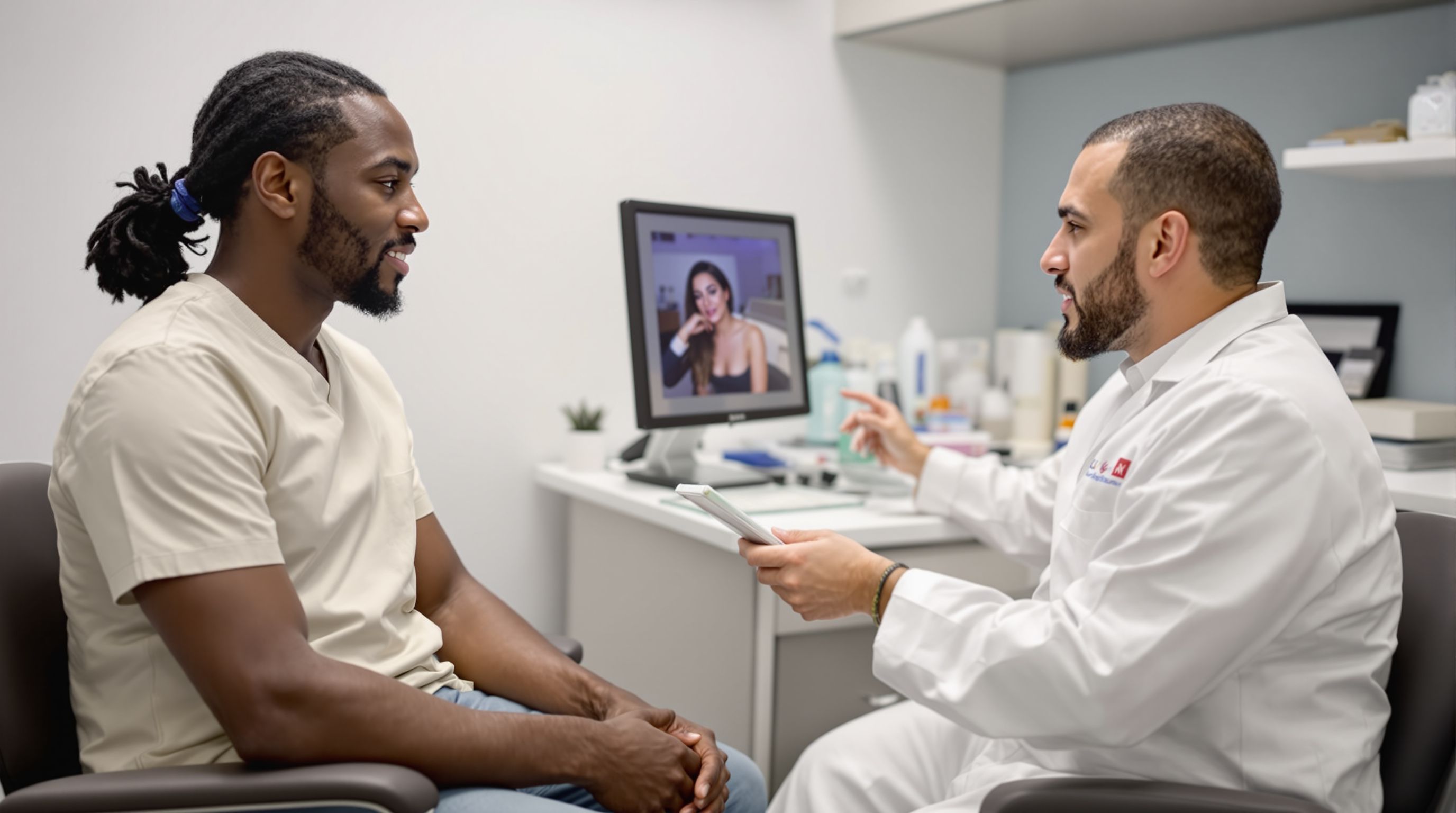
For many people living with a keloid, the first question asked in a dermatology or plastic surgery consult is simple: can you just remove it? The answer is yes, a surgeon can excise a keloid. The harder truth is what the published literature has shown for decades. When a keloid is excised and nothing else is done, recurrence rates in studies range from roughly 45 to 100 percent, and the regrown scar is sometimes larger than the original. Understanding why requires looking at what a keloid actually is, because it is not simply excess scar tissue sitting on the skin.
A keloid is a wound-healing process that never received the stop signal. Normal wound healing moves through inflammation, tissue building, and remodeling, then winds down. In keloid-prone skin, fibroblasts, the cells that produce collagen, remain abnormally activated. Research points to sustained signaling through transforming growth factor beta (TGF-beta), particularly the TGF-beta 1 and 2 pathways, which keeps fibroblasts producing thick, disorganized collagen long after the wound has closed. Keloid fibroblasts also show reduced apoptosis, meaning they resist the programmed cell death that normally clears out builder cells when the job is done.
Now consider what excision does. Surgery removes the visible collagen mass, but it also creates a brand new wound in skin that has already demonstrated it heals abnormally. The genetic and cellular predisposition does not come out with the specimen. The fresh incision triggers a new inflammatory cascade, the same overactive fibroblast population responds, and the cycle restarts, often with more surface area involved than before. This is why excision alone is widely described in the clinical literature as inadequate for most keloids rather than as a definitive cure.
Mechanical tension makes this worse. Keloids cluster in high-tension zones: the chest, shoulders, upper back, and jawline. Skin tension activates mechanotransduction pathways in fibroblasts, effectively telling them the wound is still under stress and more collagen is needed. A surgical wound under tension in a keloid-prone patient is close to a worst-case scenario. This is also why earlobe keloids, which sit in a low-tension area, tend to respond better to excision-based approaches than chest keloids do.
So what actually changes the math? The consistent theme across treatment research is combination therapy: pair the excision with something that interrupts the renewed healing response.
Corticosteroid injection after excision. Intralesional triamcinolone injected into the wound edges at surgery and at intervals afterward suppresses inflammation and reduces fibroblast collagen production. Studies of excision plus serial steroid injection generally report recurrence rates falling to roughly 15 to 50 percent, a substantial improvement over surgery alone, though results vary by site and protocol adherence.
Postoperative radiotherapy. Low-dose superficial radiation delivered within 24 to 72 hours of excision targets the rapidly dividing fibroblasts before they can rebuild the keloid. Published series report recurrence rates commonly in the 10 to 30 percent range, among the lowest of any approach. The tradeoff is access, cost, and a small but real long-term consideration around radiation exposure that patients should discuss candidly with their care team, especially for keloids near the thyroid or breast in younger patients.
Pressure therapy. For earlobe keloids specifically, custom pressure earrings worn for months after excision appear to work by inducing local tissue hypoxia and reducing collagen synthesis. Compliance is the limiting factor, since the devices typically need to be worn many hours a day for six months or longer.
Adjuncts under study. Intralesional 5-fluorouracil, alone or mixed with steroid, inhibits fibroblast proliferation and has shown promise in trials. Silicone sheeting after excision is low-risk and may help through hydration and occlusion of the healing wound, though its effect size as a standalone recurrence-prevention tool is modest.
What this means practically. If a clinician offers excision with no follow-up plan, that is a reasonable moment to ask questions. A credible surgical plan for a keloid almost always includes a named adjuvant strategy, a schedule, and a discussion of recurrence odds for that specific body site. Patients should also expect a monitoring window of at least a year, since recurrences frequently appear within the first 12 months but can emerge later.
It is also worth saying clearly: recurrence after excision is not a sign the patient did something wrong. It reflects underlying biology, including genetic factors that are more common in people of African, Asian, and Hispanic ancestry and in those with a family history of keloids.
The takeaway is not that surgery is useless. It is that surgery is one step in a protocol, not a cure on its own. The keloids that stay gone are usually the ones where the excision was immediately followed by a deliberate plan to shut down the healing response that created the keloid in the first place.
Related reading: Why Keloids Grow Where They Do: The Body's Tension Map.
Further reading: Analysis of Therapeutic Interventions for Hypertrophic and Keloid Scarring: A Systematic Review (J Drugs Dermatol 2025); Keloid management: a review of treatment modalities (Ital J Dermatol Venerol 2025); Pathogenesis, attenuation, and treatment strategies for keloid management (Tissue Cell 2025).